
Corneal Services at Rameshwaram Netralaya
Comprehensive care for corneal conditions, dry eye treatment, and advanced diagnostics to preserve vision clarity
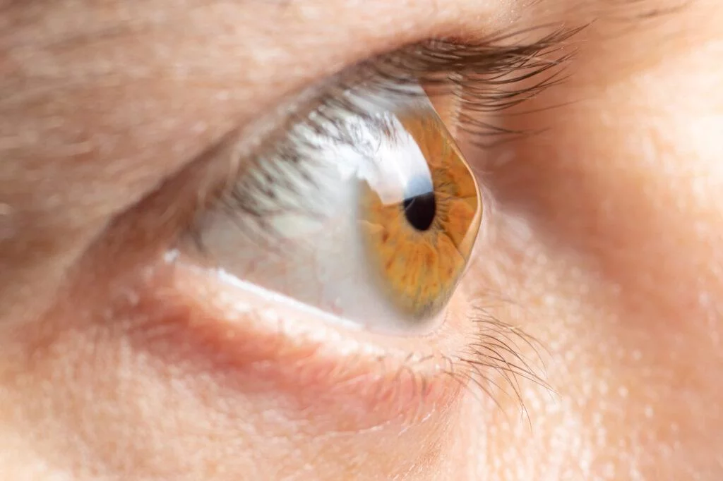
Corneal services at Rameshwaram Netralaya focus on the clear front surface of the eye, which plays a crucial role in vision clarity and eye health. Conditions affecting the cornea can cause pain, blurred vision, light sensitivity and even vision loss if untreated.
We provide comprehensive evaluation and specialized treatments to preserve corneal health and visual function, using state-of-the-art diagnostic technology and evidence-based treatment approaches.
Corneal Topography
Advanced Surface MappingCorneal topography creates a detailed, color‑coded map of the cornea's surface curvature, elevation and shape, providing essential information for diagnosis, treatment planning and monitoring. This non‑invasive test uses computerized imaging to detect subtle irregularities that standard eye exams cannot identify.
The Topography Procedure
5‑10 minutesPreparation
Patient sits comfortably while the machine captures multiple images of the corneal surface. No contact or anesthesia required.
Imaging
Advanced cameras capture thousands of data points to create a detailed 3D map of the cornea's curvature and elevation.
Analysis
Computer software analyzes the data and generates color‑coded maps that highlight irregularities and abnormalities.
Results
Immediate results available for discussion with your cornea specialist to guide personalized treatment decisions.
What Corneal Topography Reveals
Early Keratoconus Detection
Identifies corneal thinning and bulging even before symptoms appear, enabling early intervention with corneal cross‑linking.
Astigmatism Analysis
Maps astigmatism patterns and irregular astigmatism for precise glasses or contact lens prescriptions.
LASIK Candidacy Assessment
Identifies unsuitable corneal shapes or patterns that may increase LASIK risks, ensuring patient safety.
Contact Lens Fitting
Optimizes contact lens fitting, especially for irregular corneas requiring custom‑designed lenses.
Post‑Surgical Monitoring
Tracks corneal healing and shape changes after LASIK, corneal transplants, or other procedures.
Disease Progression Tracking
Monitors changes in corneal shape over time for conditions like keratoconus, pellucid marginal degeneration, etc.
Dry Eye Treatment
Comprehensive Tear Film ManagementDry eye occurs when the eyes don't produce enough tears or the tears evaporate too quickly, leading to irritation, burning, redness and fluctuating vision. At Rameshwaram Netralaya, dry eye management follows a step‑wise approach based on the severity and underlying cause.
Comprehensive Dry Eye Evaluation
Tear Film Analysis
Assessment of tear break‑up time, quantity, and quality using specialized dyes and imaging technology.
Corneal Topography
Detects surface irregularities caused by chronic dryness and monitors treatment effectiveness.
Meibomian Gland Evaluation
Infrared imaging to assess oil‑producing glands that stabilize tears and prevent evaporation.
Ocular Surface Staining
Special dyes reveal damaged areas on the corneal surface caused by inadequate tear film.
Corneal Diseases
Comprehensive Diagnosis & ManagementThe cornea can be affected by various conditions requiring specialized diagnosis and management. Rameshwaram Netralaya provides comprehensive evaluation and treatment for both common and complex corneal disorders.
Common Corneal Conditions
Advanced Diagnostics
Treatment Approaches
Medical Therapy
- Anti‑infectives for corneal infections
- Anti‑inflammatories for inflammatory conditions
- Hypertonic saline for corneal edema
- Specialized lubricants for surface disorders
Corneal Cross‑Linking
- UV light and riboflavin to strengthen thinning corneas
- Halts progression of keratoconus
- Minimally invasive outpatient procedure
- Preserves corneal shape and vision
Amniotic Membrane Transplantation
- Biological bandage for non‑healing surface defects
- Promotes healing and reduces inflammation
- Used for persistent corneal ulcers and erosions
- Minimizes scarring and promotes epithelial growth
Specialty Contact Lenses
- Scleral lenses for irregular corneas
- Hybrid lenses combining rigid and soft materials
- Custom rigid gas permeable lenses
- Prosthetic lenses for scarred corneas
Surgical Options
- DALK (Deep Anterior Lamellar Keratoplasty)
- DMEK/DSEK endothelial transplants
- Penetrating keratoplasty (full‑thickness)
- Phototherapeutic keratectomy (PTK)
Early Intervention
- Regular monitoring of high‑risk conditions
- Preventive treatments to avoid progression
- Lifestyle modifications for corneal health
- Patient education for self‑management
Early intervention preserves corneal clarity and prevents vision‑threatening complications. Regular follow‑up is essential for managing progressive corneal conditions and ensuring optimal visual outcomes.
Corneal Emergencies
Immediate Care for Urgent ConditionsCertain corneal conditions require immediate medical attention to prevent permanent vision loss. Our corneal specialists provide 24/7 emergency care for acute corneal issues.
Immediate Attention Required
URGENT- Chemical burns to the eye
- Corneal ulcers with severe pain
- Acute angle‑closure glaucoma
- Penetrating eye injuries
- Sudden vision loss with eye pain
Seek Care Within 24 Hours
PRIORITY- Foreign body in eye not removed by rinsing
- Corneal abrasion with significant discomfort
- Red eye with sensitivity to light
- Sudden blurry vision with eye redness
- Eye pain after recent eye surgery
Dr. Umesh Singh
Chief Ophthalmologist & Cataract Surgeon
Fellowship in Cornea & External Diseases
View Full Profile →Advanced Technology & Procedures
State‑of‑the‑art equipment for precise diagnosis and treatment
Corneal Topographer
Advanced Placido‑disc and Scheimpflug imaging for detailed 3D corneal mapping and early disease detection.
Anterior Segment OCT
High‑resolution cross‑sectional imaging of corneal layers for precise measurement and pathology detection.
LipiFlow® System
Advanced thermal pulsation treatment for meibomian gland dysfunction and evaporative dry eye.
Corneal Cross‑Linking
UV‑A light and riboflavin treatment to strengthen corneas and halt keratoconus progression.
Intense Pulsed Light (IPL)
Non‑invasive treatment for ocular rosacea and inflammatory dry eye conditions.
Scleral Lens Fitting
Advanced fitting systems for specialty contact lenses that vault over irregular corneas.
Corneal Services Packages
Comprehensive care plans for corneal health
Basic Corneal Evaluation
- Complete slit‑lamp examination
- Corneal thickness measurement
- Basic tear film assessment
- Consultation with cornea specialist
- Basic treatment recommendations
Advanced Corneal Work‑up
- Complete basic evaluation
- Corneal topography & tomography
- Detailed tear film analysis
- Meibomian gland imaging
- Anterior segment OCT
- Personalized treatment plan
Dry Eye Management Package
- Initial comprehensive evaluation
- Monthly follow‑up visits
- Prescription medications included
- Artificial tear supplements
- Lifestyle modification guidance
- 3‑month package available
Insurance & Payment Options
Insurance Coverage
Most corneal treatments covered by major insurers. We assist with claim processing and documentation.
EMI Options
0% interest EMI for 6‑12 months available for surgical procedures through partner banks.
Package Discounts
Special rates for comprehensive packages and long‑term management plans.
Senior Citizen Benefits
Additional 10% discount for patients above 60 years of age.
Protect Your Corneal Health & Vision Clarity
The cornea is your eye's clear window to the world. Early detection and proper management of corneal conditions can preserve your vision and prevent complications. Our corneal specialists are here to provide personalized care using the latest diagnostic technology.