Glaucoma Treatment at Rameshwaram Netralaya
Comprehensive glaucoma care including diagnosis, monitoring, laser treatments, and advanced surgical options
Glaucoma is a major cause of irreversible blindness, but early detection and timely treatment can help prevent vision loss. At Rameshwaram Netralaya, glaucoma care focuses on early diagnosis, regular monitoring and personalized treatment to control eye pressure and protect the optic nerve.
Since glaucoma often shows no early symptoms, routine eye check-ups are essential—especially for high-risk patients—to preserve long-term vision.
Understanding Glaucoma
The Silent Thief of SightGlaucoma is a group of eye conditions in which damage to the optic nerve is usually related to elevated pressure inside the eye (intraocular pressure), leading to gradual and permanent loss of vision. The most common form, primary open‑angle glaucoma, progresses slowly and without pain, so patients often do not notice any problem until a large amount of side vision is already lost.
Types of Glaucoma We Treat
Open‑Angle Glaucoma
Most common, slow progression
Angle‑Closure Glaucoma
Emergency condition, rapid onset
Normal‑Tension Glaucoma
Damage despite normal pressure
Congenital Glaucoma
Present at birth or early childhood
Secondary Glaucoma
Due to injury, disease, or medications
Acute Angle‑Closure
Medical emergency requiring immediate treatment
Glaucoma Risk Factors
Are You At Risk?Certain factors increase the likelihood of developing glaucoma. Regular screening is especially important for individuals with one or more of these risk factors.
Age Over 40
Risk increases significantly after age 40, and continues to rise with each decade
Family History
Having a parent or sibling with glaucoma increases your risk 4‑9 times
High Eye Pressure
Elevated intraocular pressure is the most significant controllable risk factor
Severe Myopia/Hyperopia
Extreme nearsightedness or farsightedness increases glaucoma risk
Long‑term Steroid Use
Prolonged corticosteroid use can elevate eye pressure
Diabetes & Hypertension
Systemic conditions that affect blood flow to the optic nerve
Ethnicity
African, Hispanic, and Asian ancestry have higher predisposition
Previous Eye Injury
Trauma can damage drainage system and increase pressure
Emergency Warning: Acute Angle‑Closure Glaucoma
If you experience sudden eye pain, headache, nausea, vomiting, blurred vision, or halos around lights, seek immediate medical attention. This is a medical emergency that can cause permanent vision loss within hours.
Emergency Hotline: +91 98765 43210Diagnosis & Screening
Comprehensive Glaucoma Work‑upGlaucoma diagnosis requires a combination of tests rather than a single reading, because some patients have damage with normal pressure and others have high pressure without damage. At Rameshwaram Netralaya, a complete glaucoma work‑up includes multiple painless, outpatient procedures.
Tonometry
5 minMeasures intraocular pressure (IOP) - raised IOP is a major risk factor for glaucoma, but normal values do not rule out the disease.
Optic Nerve Examination
10 minThe doctor evaluates the optic nerve head for thinning, cupping and structural changes using slit‑lamp lenses and optical coherence tomography (OCT).
Visual Field Testing
15‑20 minA computerized test that maps peripheral vision to detect characteristic glaucoma‑related blind spots and monitor progression over time.
Gonioscopy
5 minA special mirrored lens is used to view the drainage angle where fluid exits the eye, helping distinguish open‑angle from angle‑closure glaucoma.
Pachymetry
2 minMeasures central corneal thickness which influences how pressure readings are interpreted and is included in overall risk assessment.
OCT & Imaging
10 minHigh‑resolution imaging of the optic nerve and retinal nerve fiber layer for precise measurement and tracking of structural changes.
Laser Treatments for Glaucoma
Minimally Invasive Pressure ControlLaser therapy is an important part of modern glaucoma management and can be used as primary treatment or when drops alone are insufficient. It's important to understand that laser does not "cure" glaucoma; it is one tool among drops and surgery to keep eye pressure within a safe range.
Selective Laser Trabeculoplasty (SLT)
Most CommonFor: Open‑angle glaucoma and ocular hypertension
Procedure: Low‑energy laser pulses target pigmented cells in the trabecular meshwork, enhancing fluid outflow
Recovery: Minimal discomfort, quick recovery, typically outpatient
Duration: 10‑15 minutes per eye
Benefits:
- Reduces or eliminates need for eye drops
- Repeatable if needed
- Minimal side effects
- Can be combined with cataract surgery
Argon Laser Trabeculoplasty (ALT)
Established TechniqueFor: Open‑angle glaucoma where SLT may not be suitable
Procedure: Thermal laser treats trabecular meshwork to increase aqueous outflow
Recovery: Quick return to normal activities
Duration: 5‑10 minutes per eye
Benefits:
- Well‑established track record
- Effective pressure reduction
- Clinic‑based procedure
- Minimal downtime
Cyclophotocoagulation
Advanced CasesFor: Advanced or refractory glaucoma cases
Procedure: Laser applied to reduce fluid production by partially treating the ciliary body
Recovery: May require more recovery time than SLT/ALT
Duration: 15‑30 minutes
Benefits:
- Effective for difficult‑to‑control glaucoma
- Can avoid more invasive surgery
- Repeatable treatment option
- Reduces medication burden
Surgical Options
When Medication & Laser Aren't EnoughIf medications and laser treatments cannot adequately control eye pressure, or if very low target pressure is needed to protect the optic nerve, glaucoma surgery may be recommended. The goal of surgery is to slow or stop further damage, not to restore vision that has already been lost.
Minimally Invasive Glaucoma Surgery (MIGS)
InnovativeMIGS procedures use tiny incisions and microscopic devices to enhance fluid outflow with less tissue disruption and faster recovery time than traditional surgery. They are often combined with cataract surgery in mild to moderate glaucoma.
Ideal For:
- Mild to moderate glaucoma
- Patients undergoing cataract surgery
- Those wanting to reduce medication use
- Patients seeking quicker recovery
Trabeculectomy
Gold StandardTrabeculectomy creates a new drainage pathway under the conjunctiva, allowing fluid to leave the eye more easily and form a small reservoir ("bleb"), thereby lowering IOP. It is a well‑established operation used in more advanced glaucoma.
Ideal For:
- Moderate to advanced glaucoma
- When target pressure is very low
- Patients with progressive damage despite treatment
- Those intolerant to medications
Glaucoma Drainage Devices
Complex CasesTube shunts and other drainage implants divert fluid from inside the eye to an external reservoir, helping control pressure when trabeculectomy is unsuitable or has failed. These surgeries are generally reserved for complex or advanced cases.
Ideal For:
- Failed trabeculectomy
- Neovascular glaucoma
- Uveitic glaucoma
- Traumatic glaucoma
- Congenital glaucoma
Important: The glaucoma specialist at Rameshwaram Netralaya will design an individual plan—using eye drops, laser, surgery or a combination—based on disease stage, age, general health and lifestyle, with the shared goal of preserving useful vision for as long as possible.
Medication Management
First‑Line TreatmentEye drops are usually the first treatment for glaucoma. They work by either reducing the amount of fluid produced in the eye or improving its outflow. Proper and consistent use is crucial for effective pressure control.
Prostaglandin Analogs
Examples: Latanoprost, Bimatoprost, Travoprost
Action: Increase fluid outflow
Frequency: Once daily at bedtime
Beta‑Blockers
Examples: Timolol, Betaxolol
Action: Reduce fluid production
Frequency: Once or twice daily
Alpha‑Agonists
Examples: Brimonidine, Apraclonidine
Action: Reduce production & increase outflow
Frequency: Twice or three times daily
Carbonic Anhydrase Inhibitors
Examples: Dorzolamide, Brinzolamide
Action: Reduce fluid production
Frequency: Twice or three times daily
Proper Eye Drop Technique
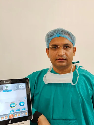
Dr. Umesh Singh
Chief Ophthalmologist & Cataract Surgeon
Treatment Success & Long‑term Monitoring
Ongoing care for lifelong vision preservation
Regular Follow‑up Visits
Every 3‑6 months for pressure checks, optic nerve evaluation, and visual field testing to monitor stability.
Progression Analysis
Advanced software compares current and previous test results to detect even subtle changes in optic nerve health.
Medication Adherence Support
Education on proper drop technique, reminder systems, and simplifying regimens to improve compliance.
Treatment Adjustment
Modifying medications, adding laser, or considering surgery if progression is detected despite treatment.
Family Screening
Encouraging and facilitating screening for family members who are at increased genetic risk.
Digital Monitoring
Remote monitoring options and digital tools to track symptoms and medication use between visits.
Treatment Packages
Comprehensive glaucoma care plans
Basic Glaucoma Screening
- Complete eye examination
- Intraocular pressure measurement
- Optic nerve evaluation
- Basic visual field screening
- Consultation with glaucoma specialist
Advanced Glaucoma Work‑up
- All basic screening tests
- Optical Coherence Tomography (OCT)
- Computerized visual field testing
- Gonioscopy & pachymetry
- Detailed risk assessment report
- 6‑month follow‑up included
SLT Laser Treatment
- Pre‑laser evaluation
- Selective Laser Trabeculoplasty
- Post‑procedure medications
- Follow‑up visits (1 week, 1 month, 3 months)
- Pressure monitoring for 6 months
Insurance & Support
Insurance Coverage
Most glaucoma treatments are covered by major insurance providers. We assist with claim processing.
EMI Options
0% interest EMI for 6‑12 months available for surgical procedures through partner banks.
Senior Citizen Discount
15% discount on all glaucoma services for patients above 60 years.
Family Screening Package
Special rates for family members of diagnosed glaucoma patients.
Don't Wait for Symptoms – Protect Your Vision Today
Glaucoma often shows no warning signs until irreversible damage has occurred. Regular screening is the only way to detect glaucoma early and preserve your vision for years to come.