
Retina & Diabetic Eye Care at Rameshwaram Netralaya
Comprehensive diagnosis and treatment for retinal diseases, diabetic retinopathy, and macular disorders
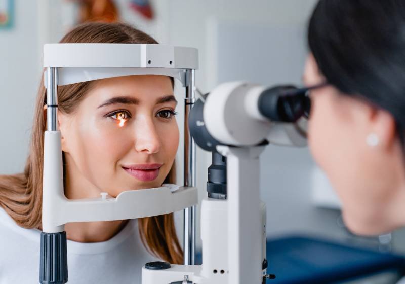
The retina is the light-sensitive layer of the eye responsible for vision, and any damage can cause permanent sight loss. Conditions like diabetes and high blood pressure can silently affect the retina, making regular retinal check-ups essential.
At Rameshwaram Netralaya, we provide comprehensive retina evaluations for diabetic and non-diabetic retinal disorders, working closely with physicians to protect and preserve your vision.
Understanding Retinal Health
The Vision‑Sensitive LayerThe retina is a delicate, light‑sensitive tissue lining the inner surface of the eye. It converts light into neural signals that travel to the brain, forming the images we see. Damage to the retina can lead to permanent vision loss, making early detection and treatment essential.
Common Retinal Conditions We Treat
Diabetic Retinopathy
Diabetes‑related retinal damage
Macular Degeneration
Age‑related central vision loss
Retinal Detachment
Emergency vision‑threatening condition
Macular Edema
Fluid accumulation in the macula
Retinal Vein Occlusion
Blockage of retinal blood vessels
Vitreous Disorders
Floaters, flashes, and hemorrhages
Diabetic Retinopathy
Diabetes‑Related Eye DamageDiabetic retinopathy is an eye disease caused by long‑term high blood sugar damaging the tiny blood vessels of the retina in people with diabetes. In the early stages, there may be no symptoms, but as the disease progresses it can lead to blurred vision, fluctuating vision, dark spots, poor night vision and even sudden, severe sight loss.
Stage 1
Mild NPDRMicroaneurysms appear as tiny bulges in retinal blood vessels
- Usually no symptoms
- Annual monitoring recommended
- Good diabetes control crucial
Stage 2
Moderate NPDRBlood vessels begin to swell and may leak fluid
- Mild blurred vision
- More frequent monitoring
- Laser may be considered
Stage 3
Severe NPDRMore blood vessels become blocked
- Significant vision changes
- High risk of progression
- Laser treatment often needed
Stage 4
Proliferative DRNew fragile blood vessels grow (neovascularization)
- High risk of bleeding
- Sudden vision loss possible
- Urgent treatment required
Other Retinal Diseases
Beyond diabetic retinopathy, several other retinal disorders can threaten central and peripheral vision and often need specialist care. Many of these conditions begin with subtle symptoms such as distortion of straight lines, patches of blurred vision, floaters, flashes of light or a curtain‑like shadow over the field of view.
Age‑related Macular Degeneration (AMD)
Progressive damage to the macula affecting central vision. Dry AMD involves thinning of macular tissue, while wet AMD involves abnormal blood vessel growth.
Retinal Vein Occlusion
Blockage of retinal veins causing bleeding, swelling, and potential vision loss. Branch retinal vein occlusion (BRVO) and central retinal vein occlusion (CRVO) are common types.
Retinal Detachment
Separation of the retina from underlying tissue – a medical emergency. Symptoms include sudden appearance of floaters, flashes of light, and shadow over vision.
Macular Disorders
Includes macular holes, epiretinal membranes, and macular edema. These conditions distort central vision and often require surgical intervention.
Intravitreal Injections
Targeted Retinal TreatmentIntravitreal injections are targeted treatments in which medicine is delivered directly into the gel (vitreous) inside the eye to act on the retina and macula. They are commonly used to treat diabetic macular edema, proliferative diabetic retinopathy, retinal vein occlusions and wet age‑related macular degeneration.
Preparation
Eye is cleaned with antiseptic solution and numbing drops are applied. Pupil may be dilated for better visualization.
Medication Administration
A tiny needle (30‑32 gauge) is used to inject medication into the vitreous cavity. The procedure takes less than 5 minutes.
Post‑Procedure Care
Eye is examined for any immediate complications. Antibiotic drops are prescribed to prevent infection.
Commonly Used Medications
Laser Therapy for Retinal Conditions
Laser treatment remains a cornerstone of retinal care, especially in diabetic retinopathy and certain other vascular retinal disorders. In diabetic eye disease, two main patterns are commonly used: focal/grid laser to seal leaking microaneurysms in or near the macula, and panretinal photocoagulation (PRP) to shrink abnormal new vessels in proliferative diabetic retinopathy.
Focal/Grid Laser
Targeted treatment for diabetic macular edema. Laser spots are placed in areas of leakage to reduce fluid accumulation and preserve central vision.
- Outpatient procedure
- Topical anesthesia
- Multiple sessions may be needed
Panretinal Photocoagulation (PRP)
Extensive laser treatment for proliferative diabetic retinopathy. Hundreds of laser spots are placed in peripheral retina to reduce oxygen demand and abnormal vessel growth.
- 2‑4 sessions typically required
- Preserves peripheral vision
- Reduces risk of severe bleeding
Barrier Laser
Creates a barrier around retinal tears to prevent progression to retinal detachment. Often used prophylactically for high‑risk tears.
- Preventive treatment
- Quick procedure
- High success rate
Advanced Diagnostic Technology
Early diagnosis using advanced tools allows timely treatment, which is crucial for preserving vision in retinal diseases. At Rameshwaram Netralaya, we utilize state‑of‑the‑art imaging technology for precise diagnosis and treatment planning.
Optical Coherence Tomography (OCT)
High‑resolution cross‑sectional imaging of the retina. Essential for detecting macular edema, epiretinal membranes, and monitoring treatment response.
Fundus Fluorescein Angiography (FFA)
Detailed imaging of retinal blood flow using fluorescent dye. Identifies leaking blood vessels, areas of non‑perfusion, and neovascularization.
Wide‑field Retinal Imaging
Captures up to 200° of the retina in a single image. Excellent for monitoring peripheral retinal changes in diabetic retinopathy.
OCT Angiography
Non‑invasive imaging of retinal blood vessels without dye injection. Ideal for patients with dye allergies or renal impairment.
Dr. Umesh Singh
Chief Ophthalmologist & Cataract Surgeon
Fellowship in Vitreo‑Retinal Surgery
View Full Profile →Prevention & Management
Proactive steps to protect your retinal health
Blood Sugar Control
Maintain HbA1c below 7% through medication, diet, and regular monitoring.
Blood Pressure Management
Keep BP below 130/80 mmHg to reduce strain on retinal blood vessels.
Healthy Diet
Consume antioxidant‑rich foods: leafy greens, fish, nuts, and colorful fruits.
Smoking Cessation
Smoking doubles the risk of AMD and accelerates diabetic retinopathy.
Regular Screenings
Annual dilated eye exams for diabetics and those with family history.
UV Protection
Wear sunglasses with UV protection to reduce oxidative stress on retina.
Treatment Packages
Comprehensive care plans for retinal conditions
Diabetic Retinopathy Screening Package
- Complete dilated eye examination
- Retinal photography (both eyes)
- Optical Coherence Tomography (OCT)
- Consultation with retina specialist
- Detailed report & follow‑up plan
Anti‑VEGF Injection Package
- Pre‑injection evaluation & OCT
- Anti‑VEGF medication (Bevacizumab)
- Intravitreal injection procedure
- Post‑procedure medications
- Follow‑up visit after 1 month
- 3‑injection package available
Laser Therapy Package
- Pre‑laser retinal evaluation
- Fluorescein angiography (if needed)
- Laser photocoagulation session
- Post‑procedure care instructions
- Follow‑up after 1‑3 months
Insurance & Payment Information
Insurance Coverage
Most retinal treatments are covered by major insurance providers. We offer direct billing facilities.
EMI Options
0% interest EMI for 6‑12 months available for treatment packages through partner banks.
Diabetic Patient Discount
Special discounted packages for diabetic patients with valid medical records.
Senior Citizen Benefits
Additional 10% discount for patients above 60 years of age.
Protect Your Vision Today
Don't wait for symptoms to appear. If you have diabetes, high blood pressure, or a family history of retinal diseases, schedule a comprehensive retinal screening at Rameshwaram Netralaya. Early detection and treatment can save your vision.